Duke University researchers have developed a new brain imaging technique that produces images 64 million times sharper than a standard MRI — and they think it could revolutionize our understanding of neurodegenerative diseases.
The challenge: The human brain is only about as large as two fists, but in that small space are 86 billion neurons, forming 100 trillion connections to one another, making the organ arguably one of the most complex systems in the universe.
While that complexity enables us to do extraordinary things, it also makes it incredibly difficult to understand what causes brain disorders, such as Parkinson’s and Alzheimer’s, and to develop effective treatments for them.
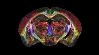
HiDiver images are 27,000 times sharper than the MRI scans typically used for mouse studies.
Better brain imaging: While mouse brains aren’t nearly as large or complex as human ones, researchers can learn a lot about neurodegenerative diseases by studying mouse models of the disorders.
The Duke-led team developed a brain imaging technique that resulted in the highest-resolution images of a whole mouse brain ever produced — these images are 27,000 times sharper than the MRI scans typically used for mouse studies.
They call the technique HiDiver (“high-dimensional integrated volume with registration”), and it builds on nearly 40 years of research at the Duke Center for In Vivo Microscopy. The details have been published in the Proceedings of the National Academy of Sciences (PNAS).
How it works: Duke’s brain imaging technique starts with an MRI scan of an anesthetized mouse, but instead of using a magnet with a strength of 1.5 to 3 Tesla — like the magnets found in MRI machines at clinics — they used a vastly more powerful 9.4 Tesla magnet.
Their scanner also includes gradient coils 100 times more powerful than those in a standard MRI and uses a high-performance computer, equivalent to 800 laptops, to process the scans.
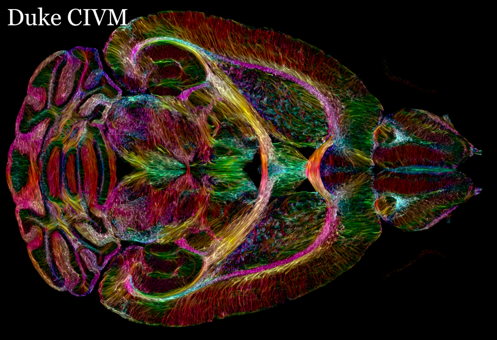
The mouse was euthanized and the brain was autopsied, stained, and scanned using light sheet microscopy — a technique that allows the researchers to label specific types of cells within the brain.
The MRI scans and light sheet images were then combined, resulting in an incredibly accurate depiction of the brain cells and the connections between them.
Looking ahead: Now that the Duke team has developed and demonstrated its new brain imaging technique, they’re eager to see researchers use it to further our understanding of aging, Parkinson’s disease, and more.
“It is something that is truly enabling,” said lead author G. Allan Johnson. “We can start looking at neurodegenerative diseases in an entirely different way.”
We’d love to hear from you! If you have a comment about this article or if you have a tip for a future Freethink story, please email us at [email protected].