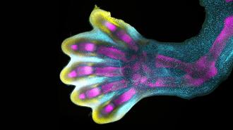
For the first time, scientists have mapped the process of limb development in human embryos down to the individual cell — and the stunning result could help prevent a common type of birth defect in the future.
The challenge: At four weeks old, the parts of a human embryo that will eventually be arms and legs are essentially buds of undifferentiated cells. By week 8, though, the limbs are well-defined, with visible fingers and toes, and we’ve never really understood how we get from point A to B.
In part, that’s because researchers have traditionally only been allowed to grow human embryos in the lab until about day 14. While that restriction is loosening, we don’t know if it’s even possible to get an embryo to develop to week 4, let alone week 8, outside a womb.
“It is like watching a sculptor at work, chiseling away at a block of marble to reveal a masterpiece.”
Hongbo Zhang
Studying young embryos inside a womb, meanwhile, is logistically tricky — at 8 weeks, an embryo is only about an inch long. Studies of animal limb development, meanwhile, might be telling us what’s going on, but we don’t know for sure since we can’t validate them.
Because we don’t fully understand the limb development process, we also don’t understand much about why it goes wrong so frequently — 1 in 500 babies is born with some significant limb abnormality, such as shortened fingers or extra toes — or how to prevent it.
What’s new? An international team of researchers, led by scientists at the Human Cell Atlas initiative, has now traced the expression of genes and differentiation of individual cells in donated fetal tissue to create the first map of human limb development.
“For the first time, we have been able to capture the remarkable process of limb development down to single cell resolution in space and time,” said senior author Sarah Teichmann.
This work led to the discovery that our fingers and toes actually don’t grow out from the clumps of limb cells we have at week 4. Instead, they form inside the buds — extra cells around them then die off to reveal the digits.
“What we reveal is a highly complex and precisely regulated process,” said senior author Hongbo Zhang. “It is like watching a sculptor at work, chiseling away at a block of marble to reveal a masterpiece. In this case, nature is the sculptor, and the result is the incredible complexity of our fingers and toes.”
Aside from exposing this remarkable process for the first time, the researchers also identified connections between common limb abnormalities and disturbances in specific genes through their study, which could be the key to preventing those abnormalities in the future.
“Our work in the Human Cell Atlas is deepening our understanding of how anatomically complex structures form, helping us uncover the genetic and cellular processes behind healthy human development, with many implications for research and healthcare,” said Teichmann.
We’d love to hear from you! If you have a comment about this article or if you have a tip for a future Freethink story, please email us at [email protected].